SARS-CoV-2 と COVID-19 に関するメモ・備忘録
COVID-19感染初期の腸内細菌叢が、後にロングCOVIDを発症するかどうかを予測可能であることが、メイヨー・クリニックの研究で判明。さらに、ロングCOVIDの症状クラスターごとに腸内細菌叢の特徴が異なることが分かりました。早期の介入が鍵となります。https://t.co/5E1HFTWXHi
— Angama (@Angama_Market) December 13, 2024
例えば、疲労症状ではエネルギー代謝に重要な Lachnospiraceae の減少、消化器症状では腸の免疫調節に関与する Prevotella の減少が関連。https://t.co/5E1HFTWXHi
— Angama (@Angama_Market) December 13, 2024
◆Gut Microbiome Signatures During Acute Infection Predict Long COVID【bioRxiv 2024年12月11日】
Abstract
Long COVID (LC), manifests in 10-30% of non-hospitalized individuals post-SARS-CoV-2 infection leading to significant morbidity. The predictive role of gut microbiome composition during acute infection in the development of LC is not well understood, partly due to the heterogeneous nature of disease. We conducted a longitudinal study of 799 outpatients tested for SARS-CoV-2 (380 positive, 419 negative) and found that individuals who later developed LC harbored distinct gut microbiome compositions during acute infection, compared with both SARS-CoV-2–positive individuals who did not develop LC and negative controls with similar symptomatology. However, the temporal changes in gut microbiome composition between the infectious (0–1 month) and post-infectious (1–2 months) phases was not different between study groups. Using machine learning, we showed that microbiome composition alone more accurately predicted LC than clinical variables. Including clinical data only marginally enhanced this prediction, suggesting that microbiome profiles during acute infection may reflect underlying health status and immune responses thus, help predicting individuals at risk for LC. Finally, we identified four LC symptom clusters, with gastrointestinal and fatigue-only groups most strongly linked to gut microbiome alterations.
SARS-CoV-2は肺、腸、心臓、腎臓、肝臓など多臓器に拡散し、各臓器内で独立して変異を続ける「区画化進化」を示すことが判明。臓器ごとに11~45の単一ヌクレオチド変異(iSNVs)を検出。これにより長期的なウイルス持続の可能性と進化の速さの要因が明らかに。https://t.co/MFajuysmWT
— Angama (@Angama_Market) December 13, 2024
◆Multi-Organ Spread and Intra-Host Diversity of SARS-CoV-2 Support Viral Persistence, Adaptation, and a Mechanism That Increases Evolvability【WILEY Online Library 2024年12月9日】
Abstract
Intra-host diversity is an intricate phenomenon related to immune evasion, antiviral resistance, and evolutionary leaps along transmission chains. SARS-CoV-2 intra-host variation has been well-evidenced from respiratory samples. However, data on systemic dissemination and diversification are relatively scarce and come from immunologically impaired patients. Here, the presence and variability of SARS-CoV-2 were assessed among 71 tissue samples obtained from multiple organs including lung, intestine, heart, kidney, and liver from 15 autopsies with positive swabs and no records of immunocompromise. The virus was detected in most organs in the majority of autopsies. All organs presented intra-host single nucleotide variants (iSNVs) with low, moderate, and high abundances. The iSNV abundances observed within different organs indicate that the virus can mutate at one host site and subsequently spread to other parts of the body. In agreement with previous data from respiratory samples, our lung samples presented no more than 10 iSNVs each. But interestingly, when analyzing different organs we were able to detect between 11 and 45 iSNVs per case. Our results indicate that SARS-CoV-2 can replicate, and evolve in a compartmentalized manner, in different body sites, which agrees with the “viral reservoir” theory. We elaborate on how compartmentalized evolution in multiple organs may contribute to SARS-CoV-2 evolving so rapidly despite the virus having a proofreading mechanism.
COVID-19感染後、自己免疫性・自己炎症性疾患のリスクが上昇することが韓国の690万人規模の調査で判明。
・関節リウマチ、クローン病、ベーチェット病、円形脱毛症など複数の疾患が関連
・初回感染後180日以上観察
感染の重症度や年齢、性別で影響が異なる。https://t.co/B00RXy9xRZ— Angama (@Angama_Market) December 17, 2024
また、感染の重症度や年齢層ごとにリスクの関連性が異なり、例えば、感染が重症だった人はベーチェット病やクローン病のリスクが顕著に上昇し、40歳未満では円形脱毛症や全身性エリテマトーデスの発症が増加しました。https://t.co/B00RXy9xRZ
— Angama (@Angama_Market) December 17, 2024
◆Long-Term Risk of Autoimmune and Autoinflammatory Connective Tissue Disorders Following COVID-19【JAMA Dermatology 2024年11月6日】
Key Points
Question Is COVID-19 infection associated with an increased long-term risk of autoimmune and autoinflammatory connective tissue disorders?
Findings This cohort study analyzing 6 912 427 participants in South Korea, including 3 145 388 with COVID-19 and 3 767 039 controls observed for more than 180 days, revealed significantly increased risks of various autoimmune and autoinflammatory connective tissue disorders following COVID-19, especially among individuals with severe COVID-19 infection, those infected with the Delta variant, and unvaccinated individuals.
Meaning These findings suggest that long-term monitoring and management of patients is crucial after COVID-19, considering demographic factors, disease severity, and vaccination status, to mitigate these risks.
Abstract
Importance Few studies have investigated the association between COVID-19 and autoimmune and autoinflammatory connective tissue disorders; however, research with long-term observation remains insufficient.
Objective To investigate the long-term risk of autoimmune and autoinflammatory diseases after COVID-19 over an extended observation period.
Design, Setting, and Participants This retrospective nationwide population-based study investigated the Korea Disease Control and Prevention Agency–COVID-19–National Health Insurance Service (K-COV-N) cohort. Individuals with confirmed COVID-19 from October 8, 2020, to December 31, 2022, and controls identified among individuals who participated in the general health examination in 2018 were included in the analysis.
Exposures Confirmed COVID-19.
Main Outcomes and Measures Incidence and risk of autoimmune and autoinflammatory connective tissue disorders in patients after COVID-19. Various covariates, such as demographic characteristics, general health data, socioeconomic status, and comorbidity profiles, were balanced using inverse probability weighting.
Results A total of 6 912 427 participants (53.6% male; mean [SD] age, 53.39 [20.13] years) consisting of 3 145 388 with COVID-19 and 3 767 039 controls with an observational period of more than 180 days were included. Alopecia areata (adjusted hazard ratio [AHR], 1.11 [95% CI, 1.07-1.15]), alopecia totalis (AHR, 1.24 [95% CI, 1.09-1.42]), vitiligo (AHR, 1.11 [95% CI, 1.04-1.19]), Behçet disease (AHR, 1.45 [95% CI, 1.20-1.74]), Crohn disease (AHR, 1.35 [95% CI, 1.14-1.60]), ulcerative colitis (AHR, 1.15 [95% CI, 1.04-1.28]), rheumatoid arthritis (AHR, 1.09 [95% CI, 1.06-1.12]), systemic lupus erythematosus (AHR, 1.14 [95% CI, 1.01-1.28]), Sjögren syndrome (AHR, 1.13 [95% CI, 1.03-1.25]), ankylosing spondylitis (AHR, 1.11 [95% CI, 1.02-1.20]), and bullous pemphigoid (AHR, 1.62 [95% CI, 1.07-2.45]) were associated with higher risk in the COVID-19 group. Subgroup analyses revealed that demographic factors, including male and female sex, age younger than 40 years, and age 40 years and older, exhibited diverse associations with the risk of autoimmune and autoinflammatory outcomes. In addition, severe COVID-19 infection requiring intensive care unit admission, the Delta period, and not being vaccinated were associated with higher risk.
Conclusions and Relevance This retrospective cohort study with an extended follow-up period found associations between COVID-19 and the long-term risk of various autoimmune and autoinflammatory connective tissue disorders. Long-term monitoring and care of patients is crucial after COVID-19, considering demographic factors, disease severity, and vaccination status, to mitigate these risks.
長引くCOVID症状の原因の一つは「迷走神経の機能不全」。この神経は自律神経のバランスや炎症抑制に関わり、機能低下が疲労や認知障害を引き起こします。研究によると「迷走神経刺激療法(VNS)」が炎症を抑え、自律神経を回復させる可能性が示唆されています。https://t.co/jXEHScm69X
— Angama (@Angama_Market) December 17, 2024
研究では「迷走神経刺激療法(VNS)」が炎症を抑え、自律神経機能を回復させることで、認知機能、疲労、睡眠の改善が報告されています。非侵襲性の「耳介迷走神経刺激」は安全で、副作用も軽微です。https://t.co/jXEHScm69X
— Angama (@Angama_Market) December 17, 2024
◆Vagal nerve stimulation for the management of long COVID symptoms【ScienceDirect 2024年11月26日】
Abstract
This review investigates the therapeutic potential of vagal nerve stimulation (VNS) in managing long COVID, a condition marked by persistent symptoms following acute SARS-CoV-2 infection. Long COVID manifests as ongoing fatigue, cognitive impairment, and autonomic dysfunction, hypothesized to arise from sustained inflammatory and neurological dysregulation. The vagus nerve, central to modulating systemic inflammation and autonomic homeostasis, represents a promising therapeutic target for symptom alleviation through VNS. A comprehensive literature search was conducted across PubMed, Scopus, and Web of Science to identify studies evaluating VNS in the context of long COVID. Preliminary evidence from small-scale pilot studies suggests VNS may attenuate systemic inflammation through activation of the cholinergic anti-inflammatory pathway (CAP), thus restoring autonomic balance and ameliorating symptoms such as fatigue, cognitive dysfunction, and anxiety. In targeting the inflammatory cascade that underlies both acute COVID-19 pathophysiology and its prolonged sequelae, VNS holds potential as an innovative intervention for persistent post-viral symptoms. While these initial findings indicate promise, current data remain limited in scope and robustness, underscoring the need for larger, controlled trials to validate the efficacy and mechanisms of VNS in long COVID management. Establishing a clearer understanding of VNS’s impact on inflammation and autonomic regulation in this context is crucial to inform clinical guidelines and therapeutic strategies for long COVID, potentially offering a targeted approach for mitigating this disabling condition.
オミクロン株は変異を重ねながらも驚異的な感染力を維持。分子動力学シミュレーションで明らかに。RBDがhACE2受容体と強固に結合し続ける仕組みとして、Q493R/KやN501Yの電荷塩橋とS375Fの安定化が判明。変異は免疫逃避と感染力のバランスを絶妙に取っている。https://t.co/LY0JAKo7Nb
— Angama (@Angama_Market) December 17, 2024
一方、K417N変異は一部の相互作用を弱めますが、これは免疫から逃れる「免疫逃避」のための調整と考えられています。https://t.co/LY0JAKo7Nb
— Angama (@Angama_Market) December 17, 2024
◆Subtle changes at the RBD/hACE2 interface during SARS-CoV2 variant evolution: a molecular dynamics study【bioRxiv 2024年12月13日】
Abstract
The SARS-CoV-2 Omicron variants present a different behavior compared to the previous variants, all particularly in respect to the Delta variant, as it seems to promote a lower morbidity although being much more contagious. In this perspective, we performed new molecular dynamics (MD) simulations of the various spike RBD/hACE2 complexes corresponding to the WT, Delta and Omicron variants (BA.1 up to BA.4/5) over 1.5 µs timescale. Then, carrying out a comprehensive analysis of residue interactions within and between the two partners, allowed us to draw the profile of each variant by using complementary methods (PairInt, hydrophobic potential, contact PCA). Main results of PairInt calculations highlighted the most involved residues in electrostatic interactions that represent a strong contribution in the binding with highly stable contacts between spike RBD and hACE2 (importance of mutated residues at positions 417, 493 and 498). In addition to the swappable arginine residues (493/498), the apolar contacts made a substantial and complementary contribution in Omicron with the detection of two hydrophobic patches, one of which was correlated with energetic contribution calculations. This study brings new highlights on the global dynamics of spike RBD/hACE2 complexes resulting from the analysis of contact networks and cross-correlation matrices able to detect subtle changes at point mutations. The results of our study are also consistent with alternative approaches such as binding free energy calculations but are more informative and sensitive to transient or low-energy interactions. Nevertheless, the energetic contributions of residues at positions 501 and 505 were in good agreement with hydrophobic interactions measurements. The contact PCA networks could identify the intramolecular incidence of the S375F mutation occurring in all Omicron variants and likely conferring them an advantage in binding stability. Collectively, these data revealed the major differences observed between WT/Delta and Omicron variants at the RBD/hACE2 interface, which may explain the greater persistence of Omicron.
Author Summary The evolution of SARS-CoV-2 was extremely rapid, leading to the global predominance of Omicron variants, despite the many mutations identified in the spike protein. Some of these were introduced to evade the immune system, but many others were located in the Receptor Binding Domain (RBD) without affecting its efficient binding to hACE2 and preserving the high infectivity of this variant. To unravel the mechanism by which this protein-protein connection remains strong or stable, it is necessary to study the different types of interactions at the atomic level and over time using molecular dynamics (MD) simulations. Indeed, in contrast to crystal or cryo-EM structures providing only a fixed image of the binding process, MD simulations have allowed to unambiguously identify the sustainability of some interactions mediated by key residues of spike RBD. This study could also highlight the interchangeable role of certain residues in compensating for a mutation, which in turn allows the virus to maintain durable binding to the host cell receptor.

先週、コロナウイルスが本質的に多くの点でただの風邪と異なることを説明しましたが、簡潔にしようとした結果いくつか誤解を生んでしまったようなので追加します。
まず以前の説明では敢えてコロナウイルス種全般に焦点を当てて説明しました。新型コロナウイルスと以前のSARSやMERSとの最も大きな違い— Angama (@Angama_Market) December 18, 2024
は、感染力です。初代やMERSはウイルス増殖メカニズムが似ていたものの、感染力が弱かったために大きく広がりませんでした。新型コロナウイルスをここまで強くしたのはスパイクプロテインの中間点に関節が生まれたためです。新型は宿主の細胞のフリンエンザイムを使ってスパイクに切り込みを入れて、
— Angama (@Angama_Market) December 18, 2024
S1とS2に柔軟性をつけることで細胞に素早く融合出来るようになりました。エボラウイルスなどはこの関節を持っていますが、コロナウイルス種の中でこの関節部を持っているものは新型だけで、どういった経緯で生まれたのかまだわかっていません。この点で「新型はただのコロナウイルスではない」と言えま
— Angama (@Angama_Market) December 18, 2024
す。また、前回説明したNSP14やORF9b、無秩序領域などの効率が他のコロナウイルスよりも飛躍的に上がっている点で長期的な病原性を高めています。たとえば狂犬病ウイルスは、感染後すぐに中枢神経系に直行し、爆発的に増殖することでほぼ100%宿主を死なせます。このウイルスは4~6時間でRNAを複製
— Angama (@Angama_Market) December 18, 2024
できます。しかし、新型コロナウイルスは複製に8~12時間要するものの、RNA自体が狂犬病よりも倍以上長いため、単位時間あたりの複製効率は致死率100%の狂犬病ウイルスもむしろ若干高いレベルです。新型コロナウイルスは中枢神経系だけではなく全身に広がるため、急性脳障害を起こすことはまれですが、
— Angama (@Angama_Market) December 18, 2024
全身の細胞にかかる負担は狂犬病ウイルスよりも大きいといえます。
もう一つ前回足りなかったのは、ヌクレオチドの説明でした。DNAやそのメッセンジャーであるRNAは、細胞に必要なタンパク質や酵素などを作る「鋳型」で、それはA、T、U、C、Gという「文字」でできています。ヌクレオチドとは— Angama (@Angama_Market) December 18, 2024
この「文字」のことで、アルファベットビスケットのようなものです。この文字で表現された文章を塩基配列と呼びます。これらの文字は細胞にとって最も重要なリソースなので、通常は厳密な監視の下で、十分なバランスと備蓄が細胞の至る所に配備してあります。しかし、よくある誤解として、
— Angama (@Angama_Market) December 18, 2024
新型コロナウイルスはDNAの配置してある細胞核で分裂するのではなく、細胞核を模倣した独自の無数の「複製複合体」を作り、その中で自己複製しています。感染した細胞の中は、細胞死が抑制されている場合数日でこの「複製複合体」で充満し、隙間が全くない状態までぱんぱんに膨らみ、細胞が内圧に
— Angama (@Angama_Market) December 18, 2024
耐えきれずに破裂するまで、これら全ての中で宿主の「文字」を使ってウイルスRNAが複製され続けます。そのため、通常は厳密に管理されている「文字」の供給が全く追いつかなくなります。これらの「複製複合体」はミトコンドリアなどと同じ二重膜でできているため、細胞内の免疫は攻撃できないのですが
— Angama (@Angama_Market) December 18, 2024
二重膜で複合体を形成するウイルスは他にいません。そのため、RNA複製をターゲットにする抗ウイルス薬は、細胞内の全ての二重膜構造に貫通しないと十分な効果を発揮できず、ウイルス増殖との競争になります。また、新型の場合はこの二重膜構造を取り巻くようにチューブ状の構造が多く確認されています
— Angama (@Angama_Market) December 18, 2024
が、この機能についてはまだほとんど分かっておらず、初代やMERSとの違いを生んでいる原因の一つの可能性もあります。複合体ではち切れそうになった細胞内では極度にストレスが増しているため、DNAに大きな損傷が生まれますが、「文字」が不足しているため、修復できず、複製もできなくなります。
— Angama (@Angama_Market) December 18, 2024
脳神経細胞は特にDNAの複製率が低いため、DNAの修復を必要としています。そのため、「文字」不足の影響は特にニューロンで大きく、免疫系の異常と重なることで、特に前帯状皮質や海馬の灰白質の神経細胞が死滅して脳容量が減少する要因の一つになります。これで生じるのがコロナウイルス長期障害による
— Angama (@Angama_Market) December 18, 2024
認知障害、神経変性疾患です。また、細胞DNAと共通して「文字」を必要とするミトコンドリアも修復が追いつかなくなることでエネルギー不足が発生します。特に三叉神経血管系でエネルギー問題が起こると片頭痛のような痛みが起こることがあります。ウイルスのRNA複製効率を担保しているタンパク質に結合
— Angama (@Angama_Market) December 18, 2024
して機能を弱めることで、ウイルスによるこの「文字」の消費を抑え、細胞が立ち直る時間を稼ぐ研究の解説が、今回の新しいレポートの要点の一つです。
— Angama (@Angama_Market) December 18, 2024
https://t.co/xplegeXFh2https://t.co/RemeY8tNvxhttps://t.co/vH7VOuG0Bihttps://t.co/ZifaOnLdsZhttps://t.co/kS84nroUjD).https://t.co/zRgMWeXfj0https://t.co/RkMOe9qWBChttps://t.co/36wCkussFP
— Angama (@Angama_Market) December 18, 2024
◆Structure, Function, and Antigenicity of the SARS-CoV-2 Spike Glycoprotein【ScienceDirect 2020年3月9日】
Summary
The emergence of SARS-CoV-2 has resulted in >90,000 infections and >3,000 deaths. Coronavirus spike (S) glycoproteins promote entry into cells and are the main target of antibodies. We show that SARS-CoV-2 S uses ACE2 to enter cells and that the receptor-binding domains of SARS-CoV-2 S and SARS-CoV S bind with similar affinities to human ACE2, correlating with the efficient spread of SARS-CoV-2 among humans. We found that the SARS-CoV-2 S glycoprotein harbors a furin cleavage site at the boundary between the S1/S2 subunits, which is processed during biogenesis and sets this virus apart from SARS-CoV and SARS-related CoVs. We determined cryo-EM structures of the SARS-CoV-2 S ectodomain trimer, providing a blueprint for the design of vaccines and inhibitors of viral entry. Finally, we demonstrate that SARS-CoV S murine polyclonal antibodies potently inhibited SARS-CoV-2 S mediated entry into cells, indicating that cross-neutralizing antibodies targeting conserved S epitopes can be elicited upon vaccination.

◆SARS-CoV-2 Cell Entry Depends on ACE2 and TMPRSS2 and Is Blocked by a Clinically Proven Protease Inhibitor【ScienceDirect 2020年3月5日】
Summary
The recent emergence of the novel, pathogenic SARS-coronavirus 2 (SARS-CoV-2) in China and its rapid national and international spread pose a global health emergency. Cell entry of coronaviruses depends on binding of the viral spike (S) proteins to cellular receptors and on S protein priming by host cell proteases. Unravelling which cellular factors are used by SARS-CoV-2 for entry might provide insights into viral transmission and reveal therapeutic targets. Here, we demonstrate that SARS-CoV-2 uses the SARS-CoV receptor ACE2 for entry and the serine protease TMPRSS2 for S protein priming. A TMPRSS2 inhibitor approved for clinical use blocked entry and might constitute a treatment option. Finally, we show that the sera from convalescent SARS patients cross-neutralized SARS-2-S-driven entry. Our results reveal important commonalities between SARS-CoV-2 and SARS-CoV infection and identify a potential target for antiviral intervention.

◆Coronavirus biology and replication: implications for SARS-CoV-2【nature reviews microbiology 2020年10月28日】
Abstract
The SARS-CoV-2 pandemic and its unprecedented global societal and economic disruptive impact has marked the third zoonotic introduction of a highly pathogenic coronavirus into the human population. Although the previous coronavirus SARS-CoV and MERS-CoV epidemics raised awareness of the need for clinically available therapeutic or preventive interventions, to date, no treatments with proven efficacy are available. The development of effective intervention strategies relies on the knowledge of molecular and cellular mechanisms of coronavirus infections, which highlights the significance of studying virus–host interactions at the molecular level to identify targets for antiviral intervention and to elucidate critical viral and host determinants that are decisive for the development of severe disease. In this Review, we summarize the first discoveries that shape our current understanding of SARS-CoV-2 infection throughout the intracellular viral life cycle and relate that to our knowledge of coronavirus biology. The elucidation of similarities and differences between SARS-CoV-2 and other coronaviruses will support future preparedness and strategies to combat coronavirus infections.
◆Host Cell and SARS-CoV-2-Associated Molecular Structures and Factors as Potential Therapeutic Targets【National Library of Medicine 2021年9月15日】
Abstract
Coronavirus disease 19 (COVID-19) is caused by an enveloped, positive-sense, single-stranded RNA virus, referred to as severe acute respiratory syndrome coronavirus 2 (SARS-CoV-2), which belongs to the realm Riboviria, order Nidovirales, family Coronaviridae, genus Betacoronavirus and the species Severe acute respiratory syndrome-related coronavirus. This viral disease is characterized by a myriad of varying symptoms, such as pyrexia, cough, hemoptysis, dyspnoea, diarrhea, muscle soreness, dysosmia, lymphopenia and dysgeusia amongst others. The virus mainly infects humans, various other mammals, avian species and some other companion livestock. SARS-CoV-2 cellular entry is primarily accomplished by molecular interaction between the virus’s spike (S) protein and the host cell surface receptor, angiotensin-converting enzyme 2 (ACE2), although other host cell-associated receptors/factors, such as neuropilin 1 (NRP-1) and neuropilin 2 (NRP-2), C-type lectin receptors (CLRs), as well as proteases such as TMPRSS2 (transmembrane serine protease 2) and furin, might also play a crucial role in infection, tropism, pathogenesis and clinical outcome. Furthermore, several structural and non-structural proteins of the virus themselves are very critical in determining the clinical outcome following infection. Considering such critical role(s) of the abovementioned host cell receptors, associated proteases/factors and virus structural/non-structural proteins (NSPs), it may be quite prudent to therapeutically target them through a multipronged clinical regimen to combat the disease.
◆Differential effect of SARS-CoV-2 infection on stress granule formation in Vero and Calu-3 cells【National Library of Medicine 2022年8月23日】
Abstract
Stress granule formation is induced by numerous environmental stressors, including sodium arsenite treatment and viral infection. Accordingly, stress granules can inhibit viral propagation and function as part of the antiviral host response to numerous viral infections. Severe acute respiratory syndrome coronavirus-2 (SARS-CoV-2) antagonizes stress granule formation, in part, via interaction between SARS-CoV-2 nucleocapsid (N) protein and Ras-GTPase-activating SH3-domain-binding protein 1 (G3BP1). However, it is unclear whether there are differential effects in different cell types. In this study, we assessed interaction between the N protein of SARS-CoV-2 S clade and G3BP1/2 in Vero and Calu-3 cells and investigated the effect of various SARS-CoV-2 strains on sodium arsenite-induced stress granule formation. Our data show that SARS-CoV-2 S clade N protein interacts with both G3BP1 and G3BP2 more strongly in Calu-3 vs. Vero cells. Consistent with this observation, infection with SARS-CoV-2 S clade induces stress granule formation in Vero but not in Calu-3 cells. However, infection with SARS-CoV-2 S clade, as well as other SARS-CoV-2 variants, inhibits sodium arsenite-induced stress granule formation in both cell lines. Taken together, our results show differential effects of SARS-CoV-2 infection on stress granule formation that is dependent on host cell type, rather than virus strain type.
Results
Effects of SARS-CoV-2 infection on stress granule formation
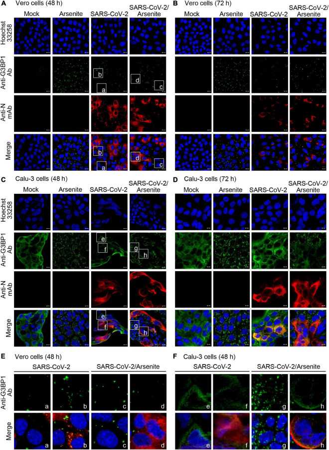
Numerous viruses, including SARS-CoV-2, are known to induce formation of stress granules within infected cells. Notably, both G3BP1 and G3BP2 are required for initiation of stress granule formation, and this process can be inhibited by the binding of viral protein to G3BP1 and G3BP2 (Matsuki et al., 2013). This suggests that SARS-CoV-2 may similarly block stress granule formation via G3BP1/G3PB2 binding. To test this hypothesis and investigate the effect of SARS-CoV-2 infection on stress granule formation, Vero and Calu-3 cells were mock-infected or infected with SARS-CoV-2 S clade stain, and stress granules were visualized by immunostaining and confocal microscopy. Sodium arsenite, a common inducer of oxidative stress that promotes stress granule formation, was used as a positive control for stress granule formation (Figure 4). We found that infection with SARS-CoV-2 S clade induces the formation of G3BP1-positive stress granules in Vero cells, but not in Calu-3 cells (Figures 4A–D). Particularly, the pattern of N protein expression and stress granule formation indicate that stress granule formation occurs in SARS-CoV-2-infected Vero cells expressing the N protein (Figure 4E, left). Conversely, we detected no stress granule formation in Calu-3 cells, irrespective of N protein expression, suggesting that the robust interaction between N and G3BP proteins effectively suppresses stress granule formation (Figure 4F, left).
◆SARS-CoV-2 N Protein Antagonizes Stress Granule Assembly and IFN Production by Interacting with G3BPs to Facilitate Viral Replication【National Library of Medicine 2022年6月2日】
Abstract
SARS-CoV-2 is the causative agent of the ongoing pandemic of coronavirus disease 2019 (COVID-19) and poses a significant threat to global health. N protein (NP), which is a major pathogenic protein among betacoronaviruses, binds to the viral RNA genome to allow viral genome packaging and viral particle release. Recent studies showed that NP antagonizes interferon (IFN) induction and mediates phase separation. Using live SARS-CoV-2 viruses, this study provides solid evidence showing that SARS-CoV-2 NP associates with G3BP1 and G3BP2 in vitro and in vivo. NPSARS-CoV-2 could efficiently suppress G3BP-mediated SG formation and potentiate viral infection by overcoming G3BP1-mediated antiviral innate immunity. G3BP1 conditional knockout mice (g3bp1fl/fL, Sftpc-Cre) exhibit significantly higher lung viral loads after SARS-CoV-2 infection than wild-type mice. Our findings contribute to the growing body of knowledge regarding the pathogenicity of NPSARS-CoV-2 and provide insight into new therapeutics targeting NPSARS-CoV-2.
IMPORTANCE In this study, by in vitro assay and live SARS-CoV-2 virus infection, we provide solid evidence that the SARS-CoV-2 NP associates with G3BP1 and G3BP2 in vitro and in vivo. NPSARS-CoV-2 could efficiently suppress G3BP-mediated SG formation and potentiate viral infection by overcoming antiviral innate immunity mediated by G3BP1 in A549 cell lines and G3BP1 conditional knockout mice (g3bp1-cKO) mice, which provide in-depth evidence showing the mechanism underlying NP-related SARS-CoV-2 pathogenesis through G3BPs.
◆COVID-19 related cognitive, structural and functional brain changes among Italian adolescents and young adults: a multimodal longitudinal case-control study【nature translational psychiatry 2024年10月2日】
Abstract
Coronavirus disease 2019 (COVID-19) has been associated with brain functional, structural, and cognitive changes that persist months after infection. Most studies of the neurologic outcomes related to COVID-19 focus on severe infection and aging populations. Here, we investigated the neural activities underlying COVID-19 related outcomes in a case-control study of mildly infected youth enrolled in a longitudinal study in Lombardy, Italy, a global hotspot of COVID-19. All participants (13 cases, 27 controls, mean age 24 years) completed resting-state functional (fMRI), structural MRI, cognitive assessments (CANTAB spatial working memory) at baseline (pre-COVID) and follow-up (post-COVID). Using graph theory eigenvector centrality (EC) and data-driven statistical methods, we examined differences in ECdelta (i.e., the difference in EC values pre- and post-COVID-19) and Volumetricdelta (i.e., the difference in cortical volume of cortical and subcortical areas pre- and post-COVID) between COVID-19 cases and controls. We found that ECdelta significantly between COVID-19 and healthy participants in five brain regions; right intracalcarine cortex, right lingual gyrus, left hippocampus, left amygdala, left frontal orbital cortex. The left hippocampus showed a significant decrease in Volumetricdelta between groups (p = 0.041). The reduced ECdelta in the left amygdala associated with COVID-19 status mediated the association between COVID-19 and disrupted spatial working memory. Our results show persistent structural, functional and cognitive brain changes in key brain areas associated with olfaction and cognition. These results may guide treatment efforts to assess the longevity, reversibility and impact of the observed brain and cognitive changes following COVID-19.
◆Mid and long-term neurological and neuropsychiatric manifestations of post-COVID-19 syndrome: A meta-analysis【National Library of Medicine 2022年1月29日】
Abstract
Importance
Neurological and neuropsychiatric symptoms that persist or develop three months after the onset of COVID-19 pose a significant threat to the global healthcare system. These symptoms are yet to be synthesized and quantified via meta-analysis.
Objective
To determine the prevalence of neurological and neuropsychiatric symptoms reported 12 weeks (3 months) or more after acute COVID-19 onset in adults.
Data sources
A systematic search of PubMed, EMBASE, Web of Science, Google Scholar and Scopus was conducted for studies published between January 1st, 2020 and August 1st, 2021. The systematic review was guided by Preferred Reporting Items for Systematic Review and Meta-Analyses.
Study selection
Studies were included if the length of follow-up satisfied the National Institute for Healthcare Excellence (NICE) definition of post-COVID-19 syndrome (symptoms that develop or persist ≥3 months after the onset of COVID-19). Additional criteria included the reporting of neurological or neuropsychiatric symptoms in individuals with COVID-19.
Data extraction and synthesis
Two authors independently extracted data on patient characteristics, hospital and/or ICU admission, acute-phase COVID-19 symptoms, length of follow-up, and neurological and neuropsychiatric symptoms.
Main outcome(s) and measure(s)
The primary outcome was the prevalence of neurological and neuropsychiatric symptoms reported ≥3 months post onset of COVID-19. We also compared post-COVID-19 syndrome in hospitalised vs. non-hospitalised patients, with vs. without ICU admission during the acute phase of infection, and with mid-term (3 to 6 months) and long-term (>6 months) follow-up.
Results
Of 1458 articles, 18 studies, encompassing a total of 10,530 patients, were analysed. Overall prevalence for neurological post-COVID-19 symptoms were: fatigue (37%, 95% CI: 25%–48%), brain fog (32%, 10%–54%), memory issues (28%, 22%–35%), attention disorder (22%, 7%–36%), myalgia (17%, 9%–25%), anosmia (12%, 8%–16%), dysgeusia (10%, 6%–14%) and headache (15%, 4%–26%). Neuropsychiatric conditions included sleep disturbances (31%, 19%–42%), anxiety (23%, 14%–32%) and depression (17%, 10%–24%). Neuropsychiatric symptoms substantially increased in prevalence between mid- and long-term follow-up. Compared to non-hospitalised patients, patients hospitalised for acute COVID-19 had reduced frequency of anosmia, anxiety, depression, dysgeusia, fatigue, headache, myalgia, and sleep disturbance at three (or more) months post-infection. Cohorts with >20% of patients admitted to the ICU during acute COVID-19 experienced higher prevalence of fatigue, anxiety, depression, and sleep disturbances than cohorts with <20% of ICU admission. Conclusions and relevance
Fatigue, cognitive dysfunction (brain fog, memory issues, attention disorder) and sleep disturbances appear to be key features of post-COVID-19 syndrome. Psychiatric manifestations (sleep disturbances, anxiety, and depression) are common and increase significantly in prevalence over time. Randomised controlled trials are necessary to develop intervention strategy to reduce disease burden.